
What is mole mapping?
Mole mapping is a screening service to enable early detection of any mole that is changing or is new. Mole mapping is a relatively new concept that involves taking top-to-toe photos of the body. The system uses a specialised magnified camera lens which enables the practitioner to zoom into any suspicious mole to check for abnormalities in appearance. We recommend anyone with moles has a yearly check up to assess mole changes and new moles. This allows our Dermatologists to detect malignant melanoma at the earliest possible stage when it is most amenable to treatment and curable with minor surgery. For anyone who is at risk of or is concerned about skin cancer, our mole mapping service is a convenient way for you and your Dermatologist to be able to monitor your moles. We recommend that anyone with moles has a yearly check up to assess mole changes and any new moles. Those at higher risk are recommended to have a mole check more regularly.
What are moles?
A mole is a small, dark, round lesion that is commonly found on the skin. Moles are known medically as melanocytic naevi which refers to the type of cells found in moles, the melanocytes. These cells produce a dark brown/black pigment (melanin) that is responsible for the dark colour of moles as well as hair and the colour of the iris in the eye. Most moles are harmless and do not cause any problems. Some moles are present from birth and others will develop during childhood and adult life. Moles can be found anywhere on the body.
What can I expect on the day of the mole mapping procedure?
On average the mole mapping service takes between 30 minutes to an hour to complete. By the end of your appointment a comprehensive ‘map’ of your moles will be created and saved in our system ready for future reference.
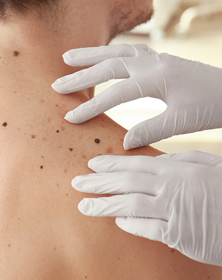
On the day of your appointment the Dermatologist will run through the procedure with you and ask if you have any questions or concerns about any particular moles. You should inform the Dermatologist if there has been any recent change in colour, size, texture and shape of your moles. Any moles which require further examination will be checked by the Dermatologist with a dermatoscope (an illuminated hand-held lens with magnification).
Our mole mapping system here at the London Dermatology Centre combines total body mapping, dermoscopy and consistent before and after photography to enable early detection of melanoma. The Dermatologist will slide the camera up and down the machine in order to create a full body profile of all the moles on your skin. The FotoFinder Medicam HD technology allows highly magnified dermoscopy images of all your moles to be taken. This allows for both immediate analysis of individual moles to produce a risk score as well as the early detection of changes in any of your previous moles.
Your Dermatologist will then give you advice on whether you need any moles looked at further or the time frame in which you should come back for another mole map. Of course if you should notice any changes or would like to come back sooner than advised just book an appointment to see your Dermatologist.
The mole mapping service is completely non-invasive and you can return to work or your daily activities immediately after the appointment.